


Purpose of this exercise
In this exercise you will search for optimal protein crystallization conditions. You will use a vapour diffusion method in hanging drops to crystallize chicken egg white lysozyme.
Read the following papers:
Lysozyme catalyzes hydrolysis of 1,4-β-linkages between N-acetylmuramic acid and N-acetyl-D-glucosamine residues in peptidoglycan and between N-acetyl-D-glucosamine residues in chitodextrins. This catalytic activity is nonspecifically targeted to the Gram positive bacterial cell wall and related with general nonspecific organism defense. Lysozyme is present in a number of secretion such as tears, saliva, human milk and mucus. Large amounts of lysozyme can be found in egg white.
In bacteriophages, lysozyme is used to break into and lyse the host bacterial cell.
Prepare the following theoretical issues:
There are a few important biochemical properties of your protein you should know before setting up a crystallization experiment. Nowadays, the primary structure (sequence) is very often the only information you can go on. Despite its relative simplicity it can deliver a plethora of useful things.
The primary sequence of chicken egg white lysozyme is given below (LYSC_CHICK P00698 ):
      1          11         21         31         41         51
      |          |          |          |          |          |
    1                    KV FGRCELAAAM KRHGLDNYRG YSLGNWVCAA KFESNFNTQA    60
   61 TNRNTDGSTD YGILQINSRW WCNDGRTPGS RNLCNIPCSA LLSSDITASV NCAKKIVSDG   120
  121 NGMNAWVAWR NRCKGTDVQA WIRGCRL
You can calculate:
The number of tryptophan and tyrosine residues. The presence of these two amino acids endow protein with substantial absorbance at 280 nm. This allows to estimate protein concentration by spectrophotometry. You can calculate the lysozyme concentration using the equation:
A = ε · t · c / M
where: A – absorbance at λ = 280 nm; c - protein concentration [g · l-1]; t – cell path length (1 cm); ε – molar extinction coefficient [l · mol-1 · cm-1]; M – protein molar mass [g · mol-1].
The molar extinction coefficient can be predicted from the equation:ε = nW · 5690 + nY · 1280
where nW, nY, are the number of tryptophan and tyrosine residues with their molar extinction coefficients, respectively. For more accurate value you should take into account the amount of phenylalanine and cystine (why not cysteine?) residues.The number of cysteine residues. The odd number of cysteines would indicate that at least one cysteine is reduced and could be used for mercury derivative to get initial phases. You should consider the cellular location of the protein. As lysozyme is present in a number of secretion one can assume it's extracellular localization which is strongly supported by its 18 residues long signal peptide at the beginning of the sequence. (Note that the sequence starts with the residue number 19).
The number of methionine residues. Generally, more than one methionine per 100 amino acids will ensure the success of a selenomet MAD experiment.
Using the ExPasy server find ProtParam tool and calculate:
Preparation of lysozyme samples, crystallization plate and setting up crystallization experiment
| 1 | 2 | 3 | 4 | 5 | 6 | |
|---|---|---|---|---|---|---|
| (A) Protein 1 c. protein 20 mg/mL, 50 mM acetate buf. pH 4.5 |
0.6 M NaCl | 0.8 M NaCl | 1.0 M NaCl | 1.2 M NaCl | 1.4 M NaCl | 1.6 M NaCl |
| (B) Protein 2 c. protein 40 mg/mL, 50 mM acetate buf. pH 4.5 |
0.6 M NaCl | 0.8 M NaCl | 1.0 M NaCl | 1.2 M NaCl | 1.4 M NaCl | 1.6 M NaCl |
| (C) Protein 3 c. protein 20 mg/mL, 50 mM acetate buf. pH 5.0 |
0.6 M NaCl | 0.8 M NaCl | 1.0 M NaCl | 1.2 M NaCl | 1.4 M NaCl | 1.6 M NaCl |
| (D) Protein 4 c. protein 40 mg/mL, 50 mM acetate buf. pH 5.0 |
0.6 M NaCl | 0.8 M NaCl | 1.0 M NaCl | 1.2 M NaCl | 1.4 M NaCl | 1.6 M NaCl |
Examination of the crystallization experiments
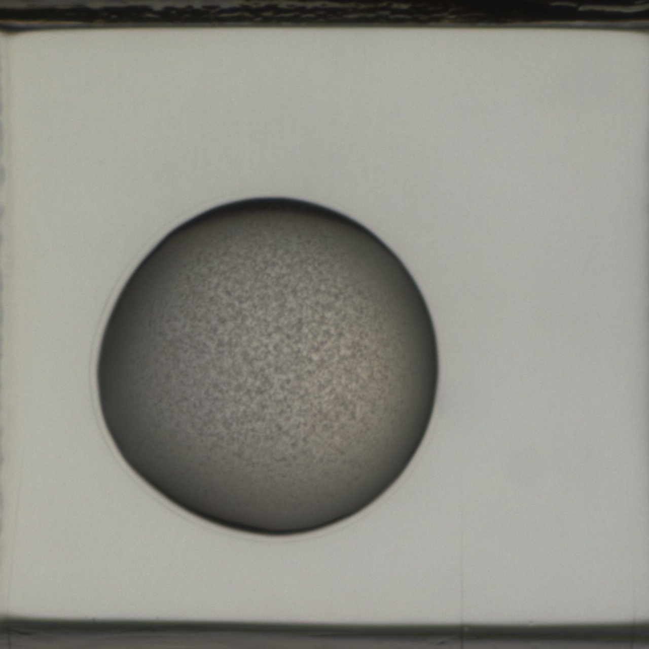
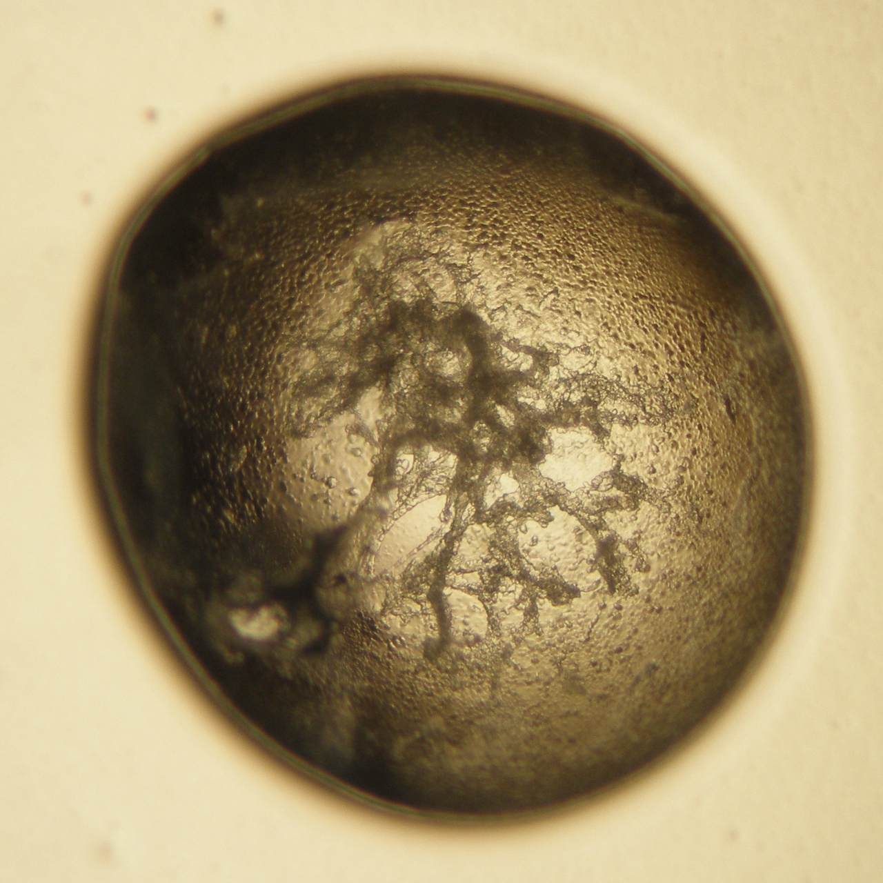
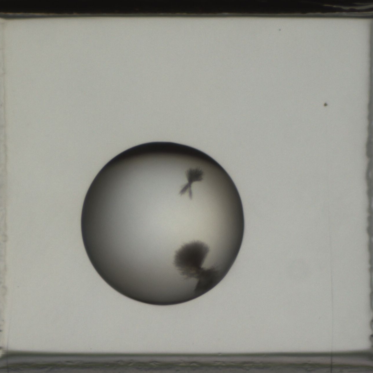
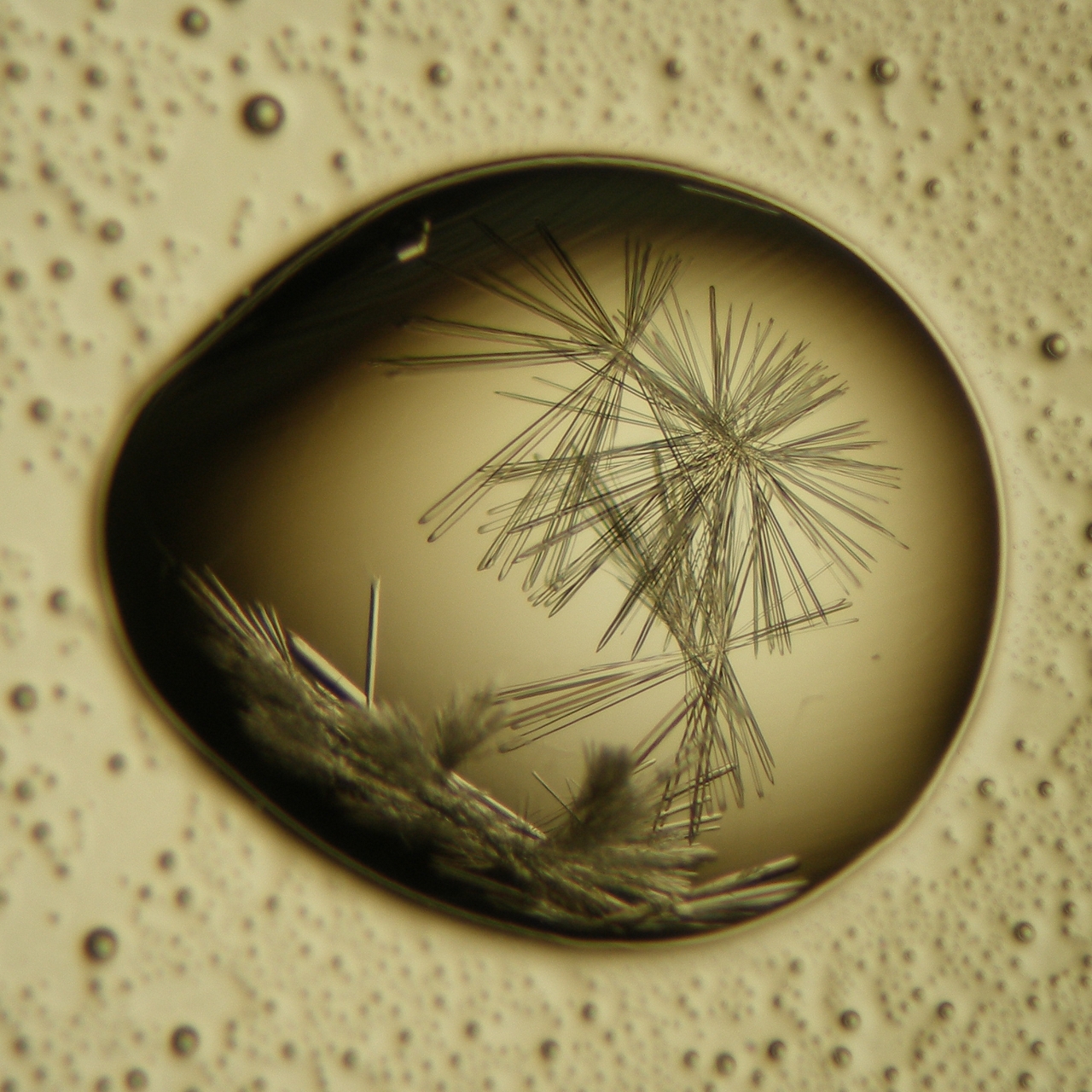
Examine the crystallization drops immediately after setup and at the beginning of the next practical. Make notes in the scoring sheet attached. You can use Johan Zeelen's numbering scheme to describe your results. The following pictures present some examples of crystallization drops you can obtain.
|
clear drop | |
|
|
non-protein particles e.g. pieces of glass, fibers from clothes |
|
mostly clear with some precipitant | |
|
skin | |
|
protein fully precipitated | |
|
|
gelatinous precipitate |
|
phase separation | |
|
spherulities | |
|
microcrystals | |
|
|
needles |
|
|
plates |
|
|
crystals |
Your Report
Last modification: Mar 14, 2014 (SW)